
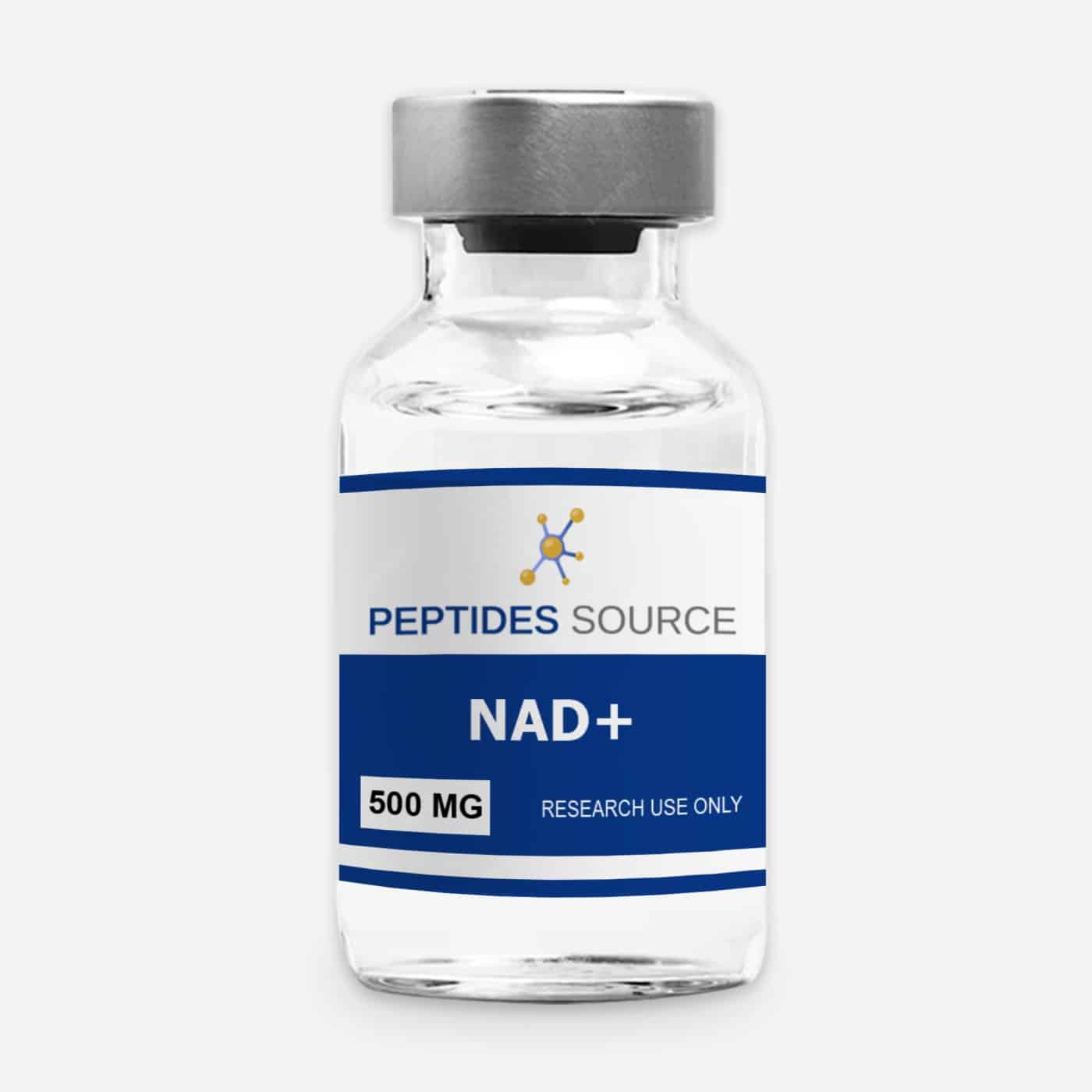

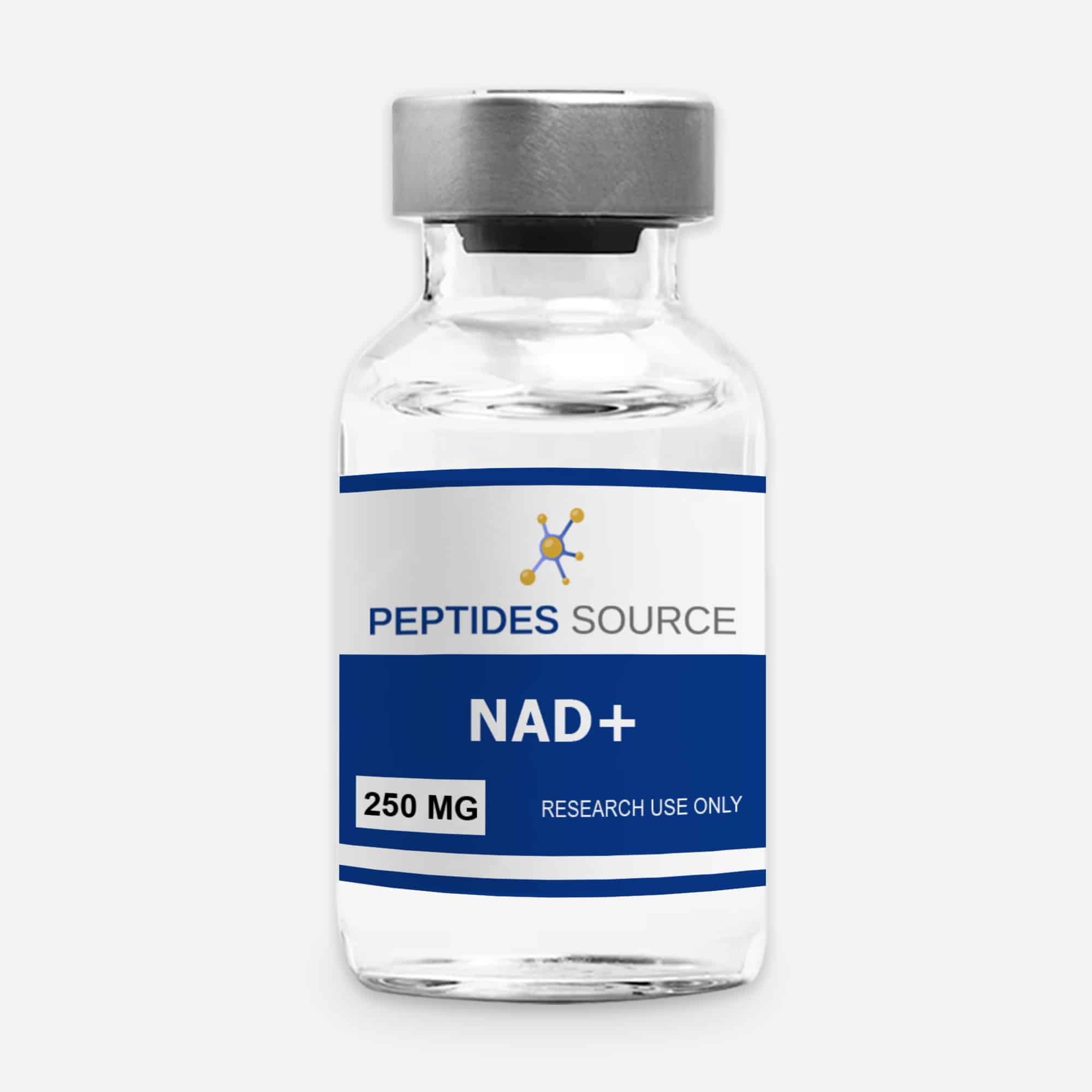
NAD+ (nicotinamide adenine dinucleotide) is an endogenous coenzyme present in every living cell, where it drives energy metabolism, supports DNA repair, and regulates mitochondrial function. Preclinical research consistently documents a decline in NAD+ levels across tissues with age, positioning it as one of the most actively studied molecules in cellular aging and metabolic biology. This profile covers what NAD+ is, why it declines with age, the mechanisms that link it to aging biology, and what preclinical research has found about interventions targeting the NAD+ pool. It is written for researchers and scientifically literate readers approaching the compound from an evidence-first perspective. NAD+ is among the most studied compounds in the cellular longevity and aging research category. All information on this page is provided for research and educational purposes only. NAD+ is supplied by Peptides Source strictly for in vitro research and laboratory use. It is not intended for human use, self-administration, or therapeutic application, and has not been approved for any medical use by the FDA or any regulatory authority.
What NAD+ Is: Biological and Chemical Profile
NAD+ (nicotinamide adenine dinucleotide, oxidized form) is a coenzyme found in all living cells. It cycles between its oxidized (NAD+) and reduced (NADH) forms, serving as an essential electron carrier in metabolic reactions — most critically in the mitochondrial electron transport chain (ETC), where it shuttles electrons from the citric acid cycle to complex I for ATP synthesis. Beyond its redox role, NAD+ functions as a substrate (consumed rather than regenerated) in three major non-redox enzymatic processes:
- Sirtuins (SIRT1–7): NAD+-dependent deacylases that regulate gene expression, mitochondrial biogenesis, stress response, and metabolic homeostasis
- PARPs (Poly-ADP-ribose polymerases): NAD+-consuming enzymes critical to DNA damage detection and repair
- CD38: A NAD+ glycohydrolase involved in calcium signaling whose activity increases substantially with age, becoming a major driver of NAD+ depletion
Key identity parameters for research-grade NAD+:
- Full name: Nicotinamide adenine dinucleotide (oxidized form)
- Molecular formula: C21H27N7O14P2
- Molecular weight: 663.43 g/mol
- CAS number: 53-84-9
- Classification: Pyridine nucleotide coenzyme / cellular energy metabolite
- Research purity standard: ≥98% by HPLC
Why NAD+ Declines With Age
The age-associated decline in NAD+ is one of the better-characterized phenomena in the biology of aging. NAD+ levels in multiple tissues — including skeletal muscle, liver, adipose tissue, brain, and skin — have been shown to decline by 40–60% between young adulthood and old age in rodent models, with parallel evidence emerging from human tissue samples. Multiple mechanisms drive this decline:
CD38 Upregulation
CD38, a NAD+ glycohydrolase expressed on immune cells and other tissue types, increases substantially with age — partly due to accumulation of senescent cells that drive CD38 expression in neighboring tissue through the senescence-associated secretory phenotype (SASP). Since CD38 is the primary consumer of NAD+ in most tissues, its age-associated increase constitutes a major drain on the NAD+ pool. Camacho-Pereira and colleagues demonstrated that CD38 knockout mice maintained higher NAD+ levels with age and were protected from some age-related metabolic decline (Cell Metabolism, 2016).
Reduced NAD+ Biosynthesis
Precursor availability for NAD+ synthesis — including nicotinamide mononucleotide (NMN) and nicotinamide riboside (NR), which feed into the salvage pathway — decreases with age due to reduced expression of key biosynthetic enzymes including NAMPT (nicotinamide phosphoribosyltransferase), the rate-limiting enzyme in the salvage pathway. This biosynthetic decline compounds the consumption-side increase from CD38 and PARP activation.
PARP Activation by DNA Damage
Accumulated DNA damage with age triggers chronic PARP activation for repair — a process that consumes NAD+ in proportion to the degree of damage. The resulting competition between DNA repair and other NAD+-dependent processes (sirtuins, mitochondrial function) has been proposed as a mechanism by which accumulated genomic damage translates into broader metabolic decline.
Sirtuins: The NAD+-Longevity Connection
The most intensively studied link between NAD+ and aging biology runs through the sirtuin family of NAD+-dependent deacylases. Sirtuins (SIRT1–7) were identified as longevity regulators through genetic studies showing that their yeast homolog (Sir2) extended lifespan when overexpressed — a finding that stimulated enormous research interest in mammalian sirtuins and their NAD+ dependence.
SIRT1 and Metabolic Regulation
SIRT1 is the most studied mammalian sirtuin and functions as a master metabolic regulator — deacetylating and thereby activating PGC-1α (which drives mitochondrial biogenesis), FOXO transcription factors (stress resistance), and other targets linked to longevity pathways. Cantó and colleagues at the Ecole Polytechnique Fédérale de Lausanne demonstrated that raising NAD+ levels by genetic or pharmacological means activated SIRT1 and improved mitochondrial function in aged mice — a landmark finding establishing the NAD+–sirtuin–mitochondria axis as a tractable research target (Cell Metabolism, 2015; PMID 26118927).
SIRT3 and Mitochondrial Protein Deacetylation
SIRT3 is the primary mitochondrial sirtuin, responsible for deacetylating and activating proteins in the electron transport chain, fatty acid oxidation, and ROS detoxification pathways. Age-associated declines in SIRT3 activity — driven by falling NAD+ — have been linked to increased mitochondrial protein acetylation and reduced oxidative phosphorylation efficiency in aged tissue. Restoring NAD+ restores SIRT3 activity in these models.
SIRT1/PARP1 Competition
A mechanistically important insight is that SIRT1 and PARP1 compete for the same NAD+ substrate. When DNA damage is high, PARP1 is prioritized and SIRT1 activity falls — a competition that may explain how genomic instability (a hallmark of aging) translates into sirtuin suppression and metabolic decline. Verdin’s review in Science formalized this framework as a unifying mechanism linking multiple hallmarks of aging through the NAD+ pool (PMID 26785480).
Preclinical Research Findings
Metabolic and Mitochondrial Function Studies
A series of studies from multiple independent research groups found that raising NAD+ levels in aged rodents — through precursor supplementation (NMN, NR) or PARP/CD38 inhibition — improved mitochondrial respiratory capacity, increased exercise performance, and reversed aspects of age-related metabolic decline. Yoshino and colleagues at Washington University School of Medicine found that NMN supplementation restored NAD+ levels and improved insulin sensitivity in aged mice on a high-fat diet, establishing the metabolic relevance of the decline (Cell Metabolism, 2011; PMID 21982712).
Muscle and Physical Function
Preclinical studies in aged rodents found that NAD+ repletion improved skeletal muscle function — including fiber composition, mitochondrial density, and ex vivo force production. Mills and colleagues demonstrated that NMN administration in aged mice improved energy metabolism and physical activity levels, with effects attributed to mitochondrial restoration downstream of SIRT1 and SIRT3 activation.
Neurological Research
NAD+ research has expanded substantially into neuroscience, given the high energy demand of neural tissue and the known role of NAD+-dependent pathways in neuronal survival. Research in models of neurodegeneration — including Alzheimer’s, Parkinson’s, and axonal degeneration — has found that NAD+ repletion or SIRT1 activation has neuroprotective effects in multiple experimental systems. Rajman, Chwalek, and Sinclair at Harvard Medical School reviewed this evidence comprehensively, identifying NAD+ as a potential target for multiple age-related neurological conditions (Cell Metabolism, 2018; PMID 29514064). This intersection with neurological aging research connects NAD+ to a broader body of work on neuroprotection and BDNF modulation that has attracted significant attention across the neuropeptide and metabolite research space.
DNA Repair and Genomic Stability
The PARP-mediated DNA repair function of NAD+ has been studied in the context of age-related genomic instability. Research has found that cells with higher NAD+ levels show faster and more complete DNA damage repair following genotoxic stress, and that aged cells with low NAD+ have compromised PARP-mediated repair — a finding consistent with the genomic instability hallmark of aging.
Cardiovascular Research
NAD+ has been studied in cardiac aging models, with findings showing that aged heart muscle has reduced NAD+ and that supplementation restores mitochondrial function and reduces markers of cardiac hypertrophy in aged rodents. Given that the heart is among the most metabolically demanding tissues and most sensitive to mitochondrial dysfunction, it has been a consistent focus of NAD+ preclinical research.
Research Purity Standards and Quality Verification
For in vitro and ex vivo NAD+ research, compound purity directly affects experimental validity. Research-grade NAD+ should meet the following specifications:
- HPLC purity: ≥98% by reverse-phase HPLC, with chromatogram data showing minimal degradation products (NADH, AMP, nicotinamide)
- Identity confirmation: UV absorbance spectrum consistent with NAD+ (λmax 260 nm in neutral solution), with mass spectrometry confirmation of MW 663.43 g/mol
- Moisture content: Low moisture is critical — NAD+ is hygroscopic and degrades readily in the presence of water
- Third-party COA: Independent laboratory documentation for each lot
Peptides Source supplies research-grade NAD+ at ≥98% HPLC purity with third-party certificate of analysis documentation for each lot. For guidance on supplier quality evaluation, see the researcher’s guide to selecting a reliable research peptide source.
Laboratory Preparation and Handling
Reconstitution and Preparation
NAD+ is typically prepared as an aqueous stock solution in sterile water or phosphate buffer (pH 7.0–7.4). Unlike most research peptides, NAD+ degrades relatively rapidly in solution — particularly at alkaline pH and elevated temperatures — so preparation of fresh working solutions immediately before use is strongly recommended for time-sensitive assays. For cell culture applications, NAD+ is typically added to media at concentrations ranging from 0.1–5 mM depending on the experimental context. Researchers should consult the standard reconstitution protocol for general lyophilized compound preparation guidance, and adapt specifically to NAD+’s pH and temperature sensitivity. For multi-use preparations requiring extended shelf life, a suitable research diluent may be appropriate, though researchers should verify that benzyl alcohol does not interfere with their specific assay system.
Storage Recommendations
- Lyophilized (unreconstituted): Store at −20°C in a sealed, desiccated container. NAD+ is highly hygroscopic — moisture exposure accelerates degradation. Keep desiccant in the storage container.
- Aqueous stock solutions: Prepare at neutral pH (7.0–7.4); store at −80°C in single-use aliquots. Avoid repeated freeze-thaw cycles.
- Working solutions: Prepare fresh immediately before use. Do not store diluted working solutions at room temperature.
- Light sensitivity: NAD+ degrades under UV exposure; store and handle in reduced-light conditions where possible.
Limitations and Open Research Questions
- Precursor vs. direct supplementation: Most in vivo NAD+ research uses precursors (NMN, NR) rather than NAD+ directly, due to NAD+’s limited cell membrane permeability. The relative efficacy of direct NAD+ vs. precursor approaches in different tissue contexts remains an active research question.
- Tissue specificity: NAD+ effects vary substantially by tissue, cell type, and baseline NAD+ status. Findings in one tissue system do not necessarily generalize, and translation across model systems requires caution.
- Route of administration in vivo: NAD+ bioavailability via different administration routes in whole-animal models is still being characterized. Oral NAD+ is largely degraded before absorption; parenteral routes are more commonly used in preclinical research.
- Species differences: NAD+ metabolism differs between mice, rats, and humans at the level of key biosynthetic enzymes. Preclinical findings, while suggestive, require independent translation to human biology.
- Sirtuin selectivity: NAD+ repletion activates all seven sirtuins to varying degrees; effects attributed to specific sirtuin activation require genetic or pharmacological tools to disambiguate.
Frequently Asked Questions: NAD+ Research
What is NAD+ and why does it matter in aging research?
NAD+ (nicotinamide adenine dinucleotide) is a coenzyme essential to mitochondrial energy metabolism, DNA repair (via PARPs), and longevity regulation (via sirtuins). Its levels decline 40–60% with age across multiple tissues, and this decline has been mechanistically linked to reduced mitochondrial function, impaired DNA repair, and sirtuin suppression — making it a central target in geroscience and aging research.
How does NAD+ activate sirtuins?
Sirtuins (SIRT1–7) are NAD+-dependent deacylases that require NAD+ as a stoichiometric substrate — consuming it in the course of removing acetyl groups from target proteins. When NAD+ is abundant, sirtuin activity is high; when NAD+ declines (as with age), sirtuin activity falls proportionally. Raising NAD+ levels in aged tissues restores sirtuin activity and the downstream metabolic benefits associated with it.
What is the role of CD38 in NAD+ decline with aging?
CD38 is a NAD+ glycohydrolase whose expression increases with age, largely driven by accumulation of senescent cells. It becomes the dominant consumer of NAD+ in aged tissues, creating a depletion dynamic that is independent of and additive to the PARP-mediated consumption from DNA damage. CD38 knockout mice maintain higher NAD+ with age and show improved metabolic function — establishing CD38 as a causative driver of age-related NAD+ decline.
What research models are used to study NAD+ biology?
Common approaches include: enzymatic cycling assays and HPLC/mass spectrometry for NAD+ quantification; Seahorse metabolic flux analysis for mitochondrial respiratory capacity; muscle fiber preparations for ex vivo contractile function; in vivo metabolic phenotyping (glucose tolerance, insulin sensitivity); and genetic models (CD38 KO, SIRT1/SIRT3 KO or overexpression) to dissect pathway contributions.
Does NAD+ cross the cell membrane in research applications?
NAD+ has limited permeability across plasma membranes under normal conditions. Most cell-based research uses extracellular NAD+ concentrations high enough to achieve intracellular effects via receptor-mediated mechanisms (CD38, P2X7 receptor) or uses precursors (NMN, NR) that enter cells via transporters (Slc12a8 for NMN; NRK1/2 for NR) and are converted intracellularly. In vitro, plasma membrane permeabilization is sometimes used to deliver NAD+ directly to the cytoplasm.
What is the difference between NAD+, NMN, and NR in research?
NAD+ is the active coenzyme. NMN (nicotinamide mononucleotide) and NR (nicotinamide riboside) are biosynthetic precursors that enter cells more efficiently and are converted to NAD+ intracellularly. Most in vivo longevity research has used NMN or NR supplementation rather than direct NAD+ due to bioavailability considerations. The relative tissue-specific efficacy of each precursor versus direct NAD+ is an ongoing area of investigation.
Is there human data on NAD+ and aging?
Several small clinical studies have examined NMN and NR supplementation in older adults, finding evidence of increased muscle NAD+ levels and some metabolic parameters with supplementation. These studies are early-phase and not powered or designed for efficacy conclusions, but provide human proof-of-concept for the preclinical finding that NAD+ precursor supplementation can raise tissue NAD+ in vivo.
What purity standard should research-grade NAD+ meet?
≥98% by HPLC (reverse-phase), with identity confirmed by UV absorbance at 260 nm and mass spectrometry verifying MW of 663.43 g/mol (CAS 53-84-9). Low moisture content is especially important — NAD+ is hygroscopic and moisture drives degradation. Third-party COA documentation is the expected verification standard.
How should NAD+ be stored for research use?
Lyophilized NAD+: −20°C in a sealed, desiccated container away from light and moisture. Aqueous stocks: −80°C in single-use aliquots at neutral pH; avoid freeze-thaw cycles. Working solutions: prepare fresh immediately before use. NAD+ degrades significantly in solution at room temperature and at alkaline pH.
Summary and Research Context
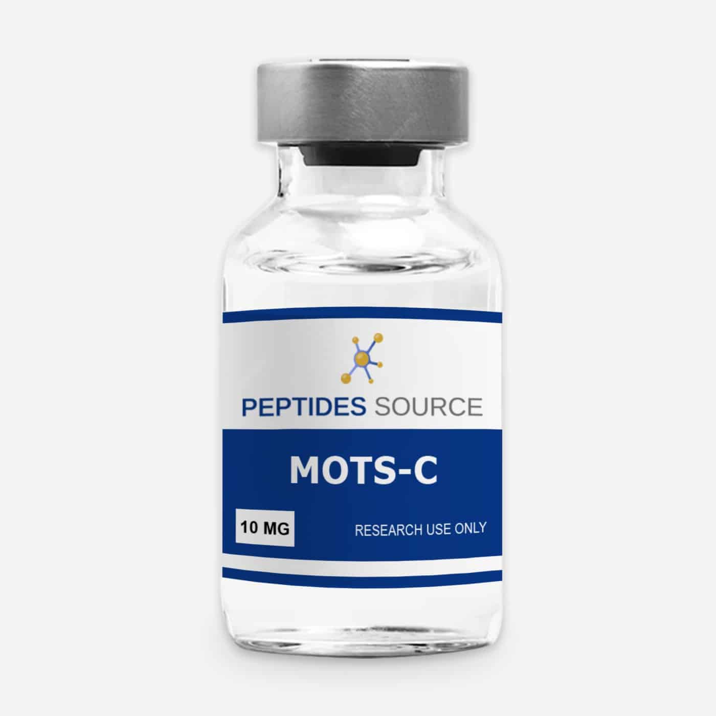
NAD+ has emerged as one of the most consequential molecules in aging biology research — not because it is a drug target in the conventional sense, but because it sits at the convergence of multiple core aging mechanisms: mitochondrial dysfunction, genomic instability, sirtuin silencing, and cellular senescence. The finding that it declines universally with age, and that preclinical restoration of NAD+ reverses or attenuates aspects of aged tissue function across multiple organ systems, has made it a central focus of geroscience research. The open questions — around membrane permeability, tissue specificity, precursor vs. direct supplementation, and species translation — are the frontier of an active and rapidly developing research field. For researchers studying aging biology, mitochondrial function, or longevity-related pathways, NAD+ is a compound with an unusually broad and mechanistically coherent evidence base. In mitochondrial-function research, NAD+ is frequently studied alongside mitochondrially derived peptides such as MOTS-c. Peptides Source supplies research-grade NAD+ at ≥98% HPLC purity with third-party COA documentation, manufactured under cGMP/ISO-compliant conditions for qualified laboratory use.
References
- Verdin E. NAD⁺ in aging, metabolism, and neurodegeneration. Science. 2015;350(6265):1208–1213. PMID 26785480
- Cantó C, Menzies KJ, Auwerx J. NAD⁺ metabolism and the control of energy homeostasis: a balancing act between mitochondria and the nucleus. Cell Metabolism. 2015;22(1):31–65. PMID 26118927
- Yoshino J, Mills KF, Yoon MJ, Imai S. Nicotinamide mononucleotide, a key NAD⁺ intermediate, treats the pathophysiology of diet- and age-induced diabetes in mice. Cell Metabolism. 2011;14(4):528–536. PMID 21982712
- Rajman L, Chwalek K, Sinclair DA. Therapeutic potential of NAD-boosting molecules: the in vivo evidence. Cell Metabolism. 2018;27(3):529–547. PMID 29514064
- Camacho-Pereira J, Tarragó MG, Chini CCS, et al. CD38 dictates age-related NAD decline and mitochondrial dysfunction through an SIRT3-dependent mechanism. Cell Metabolism. 2016;23(6):1127–1139. PMID 27304511
- Mills KF, Yoshida S, Stein LR, et al. Long-term administration of nicotinamide mononucleotide mitigates age-associated physiological decline in mice. Cell Metabolism. 2016;24(6):795–806. PMID 28068222
- Imai SI, Guarente L. NAD+ and sirtuins in aging and disease. Trends in Cell Biology. 2014;24(8):464–471. PMID 24786309
- Horenstein AL, Chillemi A, Zaccarello G, et al. A CD38/CD203a/CD73 ectoenzymatic pathway independent of CD39 drives a novel adenosinergic loop in human T lymphocytes. Oncoimmunology. 2013;2(9):e26246. PMID 24319640
Research Use Only. NAD+ is supplied by Peptides Source strictly for in vitro research and laboratory use by qualified investigators. It is not intended for human or veterinary use, self-administration, or therapeutic application. This content is educational and does not constitute medical advice. Users are responsible for compliance with all applicable local, state, and federal regulations governing the use of research compounds.