

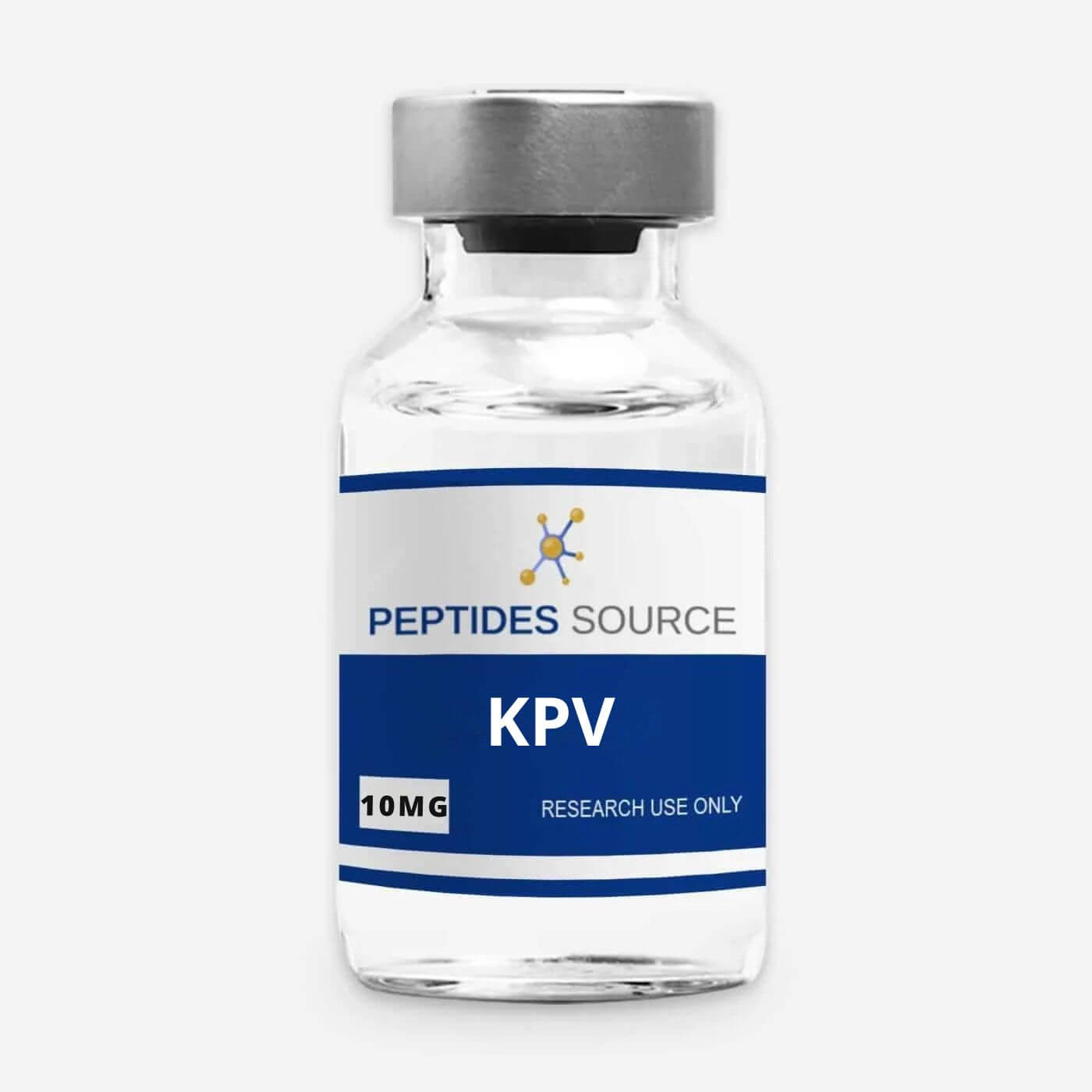
Disclaimer — For Research Use Only. This article is a scientific overview of KPV as a research compound, intended for qualified professionals studying inflammation and tissue-repair biology in vitro and in preclinical models. It is not medical or veterinary guidance. KPV offered as a research compound is not a pharmaceutical product, is not for human or animal consumption, and nothing here should be read as a description of human use, dosing, or therapeutic effect.
Key Takeaways
- KPV is a tripeptide (Lys-Pro-Val) corresponding to the C-terminal fragment of α-melanocyte-stimulating hormone (α-MSH), specifically residues 11–13.
- At just 342.43 g/mol it is one of the smallest peptides studied for anti-inflammatory mechanisms, and its small size allows cellular uptake through the di/tripeptide transporter PepT1.
- In preclinical models its anti-inflammatory activity is largely receptor-independent: once inside the cell, it interferes with the NF-κB signaling pathway, a master regulator of pro-inflammatory gene expression.
- The most cited preclinical work involves murine models of intestinal inflammation, where PepT1-mediated uptake of KPV was associated with reduced inflammatory signaling.
- For reproducible research, KPV should be characterized by ≥98% HPLC purity with a third-party certificate of analysis (COA) confirming identity and net peptide content.
What Is KPV?
KPV is a synthetic tripeptide composed of the amino acids lysine (K), proline (P), and valine (V). It is not an arbitrary sequence: it corresponds to the C-terminal three residues of α-melanocyte-stimulating hormone (α-MSH), a 13-amino-acid neuropeptide with well-documented anti-inflammatory properties in the research literature. KPV represents the minimal fragment of α-MSH that retains a substantial portion of that anti-inflammatory activity while shedding the pigmentary (melanocortin-receptor) signaling associated with the full hormone.
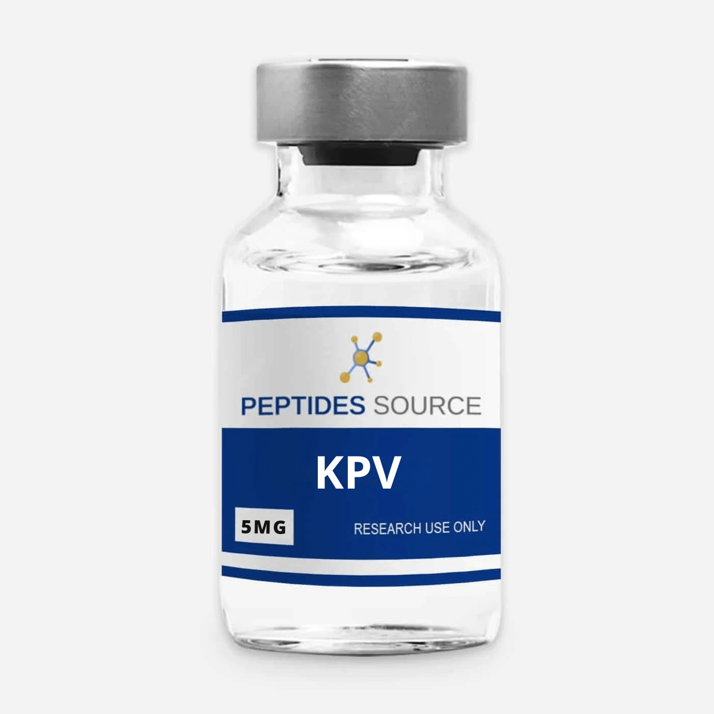
Because it is small, stable, and structurally simple, KPV is a widely studied model compound for researchers investigating inflammatory signaling and tissue-repair biology. For laboratory work it is supplied in tablet/capsule and lyophilized forms, including KPV 500mcg (100 tablets) and KPV 5mg.
Molecular Profile
KPV’s defining feature is its minimalism — three residues carrying the anti-inflammatory “message” of a much larger hormone:
| Property | Value |
|---|---|
| Peptide sequence | Lys-Pro-Val (K-P-V) |
| Parent molecule | α-MSH, residues 11–13 (C-terminal tripeptide) |
| Molecular formula (free base) | C16H30N4O4 |
| Molecular weight | 342.43 g/mol |
| PubChem CID | 125672 |
| CAS number | 67727-97-3 |
| Typical research salt form | Acetate |
(As with other research peptides supplied as salts, a certificate of analysis reports net peptide content after accounting for counter-ion and residual moisture.)
Mechanism of Action (Research Context)
KPV is studied primarily as an anti-inflammatory tripeptide, and its proposed mechanism is notable for being largely independent of the melanocortin receptors that mediate α-MSH’s classical signaling. Two features define its behavior in experimental systems:
- PepT1-mediated cellular uptake. PepT1 is a di/tripeptide transporter expressed on intestinal epithelial cells and certain immune cells, and its expression is induced during inflammation. KPV is small enough to be carried into the cell through this transporter rather than depending on a cell-surface signaling receptor.
- Intracellular interference with NF-κB. Once inside the cell, α-MSH C-terminal peptides have been shown in vitro to reach the nucleus and interfere with the NF-κB pathway — a central regulator of pro-inflammatory gene transcription. Reported effects include reduced activation of NF-κB and MAP-kinase signaling and lower secretion of pro-inflammatory cytokines.
This combination — transporter-mediated entry plus intracellular pathway interference — is what makes KPV a distinctive model compound compared with peptides that act at cell-surface receptors.
KPV and the α-MSH / Melanocortin Connection
Understanding KPV means understanding its parent. α-MSH is a melanocortin peptide best known for pigmentary signaling, but its C-terminal region carries separable anti-inflammatory and antimicrobial activity. KPV isolates that C-terminal message. Researchers comparing melanocortin-derived compounds often study KPV precisely because it lets them examine the anti-inflammatory arm of α-MSH biology without the confounding pigmentary signaling — a useful separation of function in mechanistic work. This places KPV in a different research lane from the melanocortin-receptor agonists, even though they share a common hormonal ancestor.
Preclinical Research Applications (In-Vitro / Animal Models)
In laboratory settings, KPV is used chiefly as a probe of anti-inflammatory signaling:
- Intestinal-inflammation models. The most cited work comes from murine colitis models, where PepT1-mediated uptake of KPV was associated with reduced inflammatory signaling and lower pro-inflammatory cytokine expression. This body of work established PepT1 as the route of entry underlying the effect.
- In-vitro NF-κB studies. Cell-based assays in epithelial and immune cell lines are used to characterize how α-MSH C-terminal peptides interfere with NF-κB activation.
- Structure-activity work. As the minimal active fragment of α-MSH, KPV is a reference point for studies mapping which residues carry anti-inflammatory versus pigmentary activity.
All such work assumes a qualified-researcher, in-vitro or preclinical context; none of it describes or implies human use.
Where KPV Sits Among Tissue-Repair Research Peptides
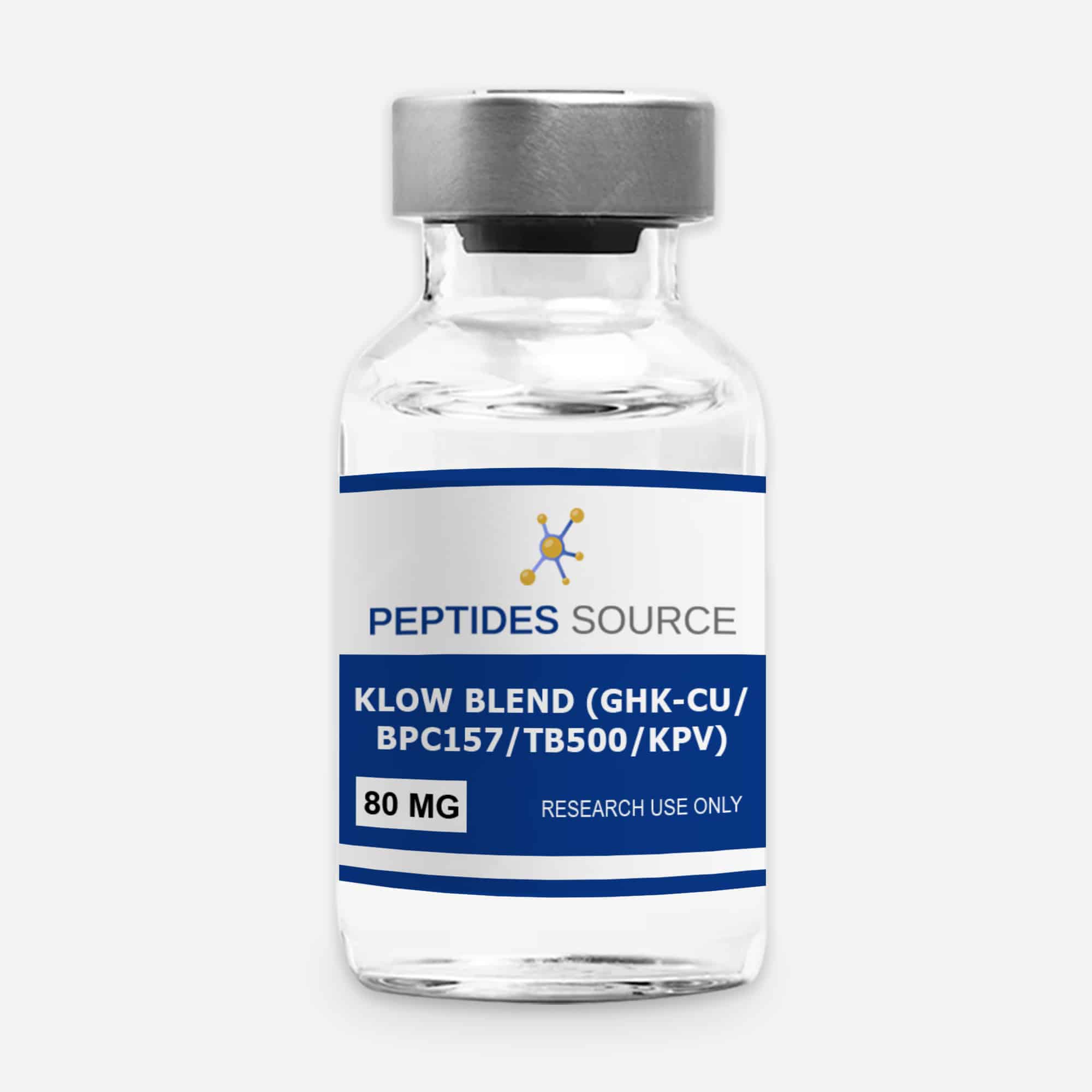
KPV is frequently studied alongside other compounds in the tissue-repair and regeneration research space, where inflammation is a central variable. It is a component of some multi-peptide research blends and is often examined next to other regeneration-focused peptides. Researchers building a tissue-repair reference set can review the broader landscape in our tissue repair research peptides overview and explore the compounds available on the Tissue Repair & Regeneration Research category page.
Handling, Reconstitution, and Quality Considerations
KPV supplied in lyophilized form requires reconstitution before use in solution-based assays; capsule and tablet formats are used where a solid form suits the research design. As a small peptide, KPV is subject to relatively rapid metabolism, so careful handling and cold storage help preserve the intact molecule for reproducible results. Our laboratory guide to reconstituting research peptides covers diluent selection, technique, and concentration math.
Reproducible work also depends on knowing exactly what is in the vial. KPV should be sourced with ≥98% HPLC purity and a third-party certificate of analysis (COA) documenting identity, mass, and net peptide content. Peptides Source manufactures to cGMP/ISO standards in the USA and provides third-party COAs across its research catalog.
Frequently Asked Questions
What is KPV?
KPV is a tripeptide made of lysine, proline, and valine (Lys-Pro-Val). It corresponds to the C-terminal three residues (positions 11–13) of α-melanocyte-stimulating hormone (α-MSH) and is studied as an anti-inflammatory research compound.
What does KPV do in research models?
In preclinical and in-vitro models, KPV is studied for its anti-inflammatory activity. It is taken into cells via the PepT1 transporter and then interferes with the NF-κB signaling pathway, which reduces pro-inflammatory signaling in these experimental systems.
How is KPV related to α-MSH?
KPV is the C-terminal tripeptide fragment of α-MSH. It retains much of the parent hormone’s anti-inflammatory activity while lacking the pigmentary (melanocortin-receptor) signaling of the full 13-amino-acid hormone, which is why it is useful for isolating that mechanism in research.
Is KPV’s anti-inflammatory effect receptor-dependent?
Research suggests the effect is largely receptor-independent. Rather than signaling through a cell-surface receptor, KPV enters the cell via the PepT1 transporter and acts intracellularly on the NF-κB pathway — a mechanism distinct from that of cell-surface receptor agonists.
How should KPV purity be verified?
Identity and purity should be confirmed by reversed-phase HPLC (target ≥98%) with mass-spectrometric confirmation of molecular weight, documented on a third-party certificate of analysis. Consistent analytical characterization is what makes research results reproducible.
Disclaimer — For Research Use Only. The information above is provided solely for in-vitro and preclinical research context. KPV offered as a research compound is not a pharmaceutical product, is not intended for human or animal consumption, and none of the molecular, mechanistic, or preclinical information here describes or implies human use, dosing, or therapeutic effect.
References
- PubChem, Compound Summary: Lys-Pro-Val / KPV (CID 125672), National Center for Biotechnology Information.
- Dalmasso G, et al. PepT1-mediated tripeptide KPV uptake reduces intestinal inflammation. Gastroenterology. 2008;134(1):166–78. PubMed (National Library of Medicine).
- Wu W, et al. Critical role of PepT1 in colitis-associated cancer and therapeutic benefits of the anti-inflammatory PepT1-mediated tripeptide KPV in a murine model. Cell Mol Gastroenterol Hepatol. 2016.
- α-Melanocyte-stimulating hormone 10–13 (GKPV) inhibits tumor-necrosis-factor-α-stimulated NF-κB activity. Peptides (ScienceDirect).